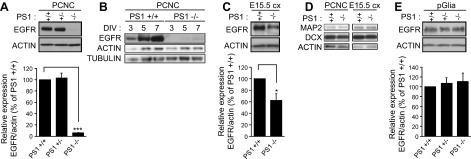
Figure 3.

PS1 positively correlates with cellular levels of neuronal EGFR in vitro and in vivo. A) (Top) Lysates from WT, PS1+/−, or PS1−/− PCNC grown in 6-well plates were prepared at 9 DIV and probed on WBs for the indicated proteins. (Bottom) Densitometric analysis of the relative amounts of EGFR in PCNC is expressed as ratio of EGFR to actin. B) Lysates from WT (+/+) or PS1−/− PCNC grown as above were prepared at 3, 5, or 7 DIV and probed on WBs for indicated proteins. C) (Top) Lysates from embryonic brain cortex (E15.5 cx) from WT or PS1 homozygous KO were prepared as described in the Materials and Methods. (Bottom) Densitometric analysis of the relative amounts of EGFR in embryonic cortices is expressed as ratio of EGFR to actin. D) (Left) Lysates from WT or PS1−/− PCNC grown as above were prepared at 9 DIV and probed on WBs for the indicated proteins. (Right) Lysates from WT and PS1−/− embryonic brain cortex (E15.5 cx) were prepared and probed on WBs for indicated proteins. E) (Top) Primary glial cultures (pGlia) from WT, PS1 heterozygous, or homozygous KO were obtained as described in the Materials and Methods. Cells were cultured in 6-well plates. When cells reached about 80% confluence, lysates were collected and assayed on WBs for the proteins indicated. (Bottom) Densitometric analysis of the relative amounts of EGFR in primary glial cultures is expressed as ratio of EGFR to actin. Data were respectively obtained from at least 4 separate experiments. *P < 0.05; ***P < 0.001 (paired Student’s t test).
