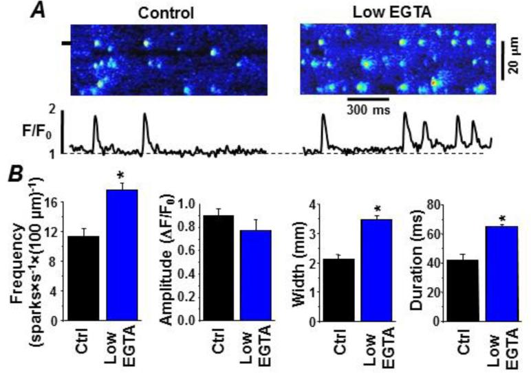
Figure 4. Low cytosolic buffer power enhances spark activity in permeabilized ventricular myocytes.
Sparks were detected as local changes in cytosolic Fluo-4 fluorescence. A. Representative sparks in the control and BAPTA-containing cytosolic recording solutions. The control cytosolic solution had 0.36 mM EGTA (150 nM free [Ca2+]). The low EGTA cytosolic solution had 0.12 mM EGTA (150 nM free [Ca2+]). Line scan images are shown at top. At the bottom are fluorescence profiles at the point marked on the line scan images (see left margin). B. Summary results of various measured spark parameters in the control and low EGTA test conditions.

