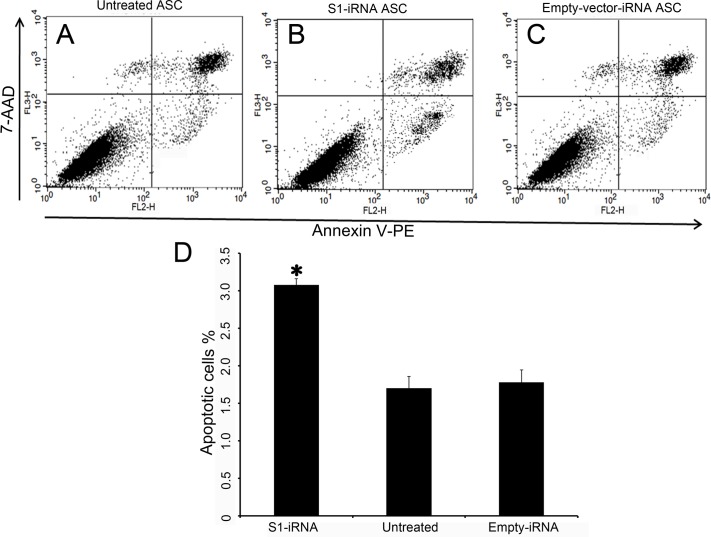
Fig 7. Effects of p21 knockdown on apoptosis of the ASCs.
Detection of cell apoptosis by Annexin V-PE/7-AAD staining. A-C: Annexin V-PE was read in FL2 and plotted on the x-axis. 7-AAD was read in FL3 and plotted on the y-axis. Living cells were located in the lower left quarter (both annexin and 7AAD negative), apoptotic cells were in the lower right quarter (annexin positive, 7AAD negative) and the dead cells (and late apoptosis) were in the top right quarter (both annexin and 7AAD positive). A, Untreated ASCs. B, S1-iRNA ASCs. C, Empty-vector-iRNA ASCs. D, Comparisons of the apoptotic rate between the S1-iRNA ASCs, the empty-vector-iRNA ASCs and the untreated ASCs. Note that the rate of apoptosis in the S1-iRNA ASCs was significantly increased compared to the untreated ASCs and the empty-vector-iRNA ASCs (P<0.05).

