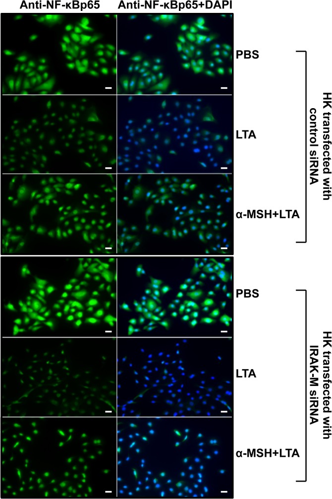
Fig 5. Inhibitory effect of IRAK-M siRNA on α-MSH-suppressed HK NF-κB nuclear translocation.
After HK cells were transiently transfected with IRAK-M siRNA or control siRNA prior to LTA treatment, NF-κB cellular localization was determined by immunofluorescent staining using a specific anti-human NF-κBp65 antibody 30 minutes after LTA treatment in the presence or absence of α-MSH. The left column is the FITC fluorescent signal indicative of NF-κB, the center column is the DAPI nuclear stain, and the right column is the merged image demonstrating nuclear or cytoplasmic of NF-κB localization. PBS-treated HK cells served as the negative control. Bars = 20 μm.

