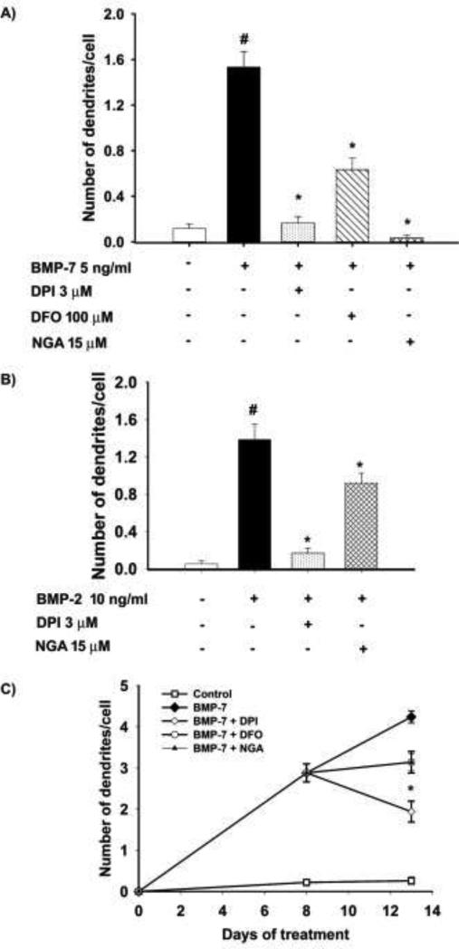
Figure 3. Antioxidants inhibited dendritic growth induced by multiple members of the BMP subfamily and caused inhibition or retraction of existing dendrites.
Sympathetic neurons cultured from perinatal rat SCG were treated for 3 d with (A) BMP-7 at a submaximal concentration (5 ng/ml) or (B) BMP-2 at a submaximal concentration (5 ng/ml) in the absence or presence of DPI (3 μM), DFO (100 μM) or NGA (15 μM). The number of dendrites per cell was quantified in cultures immunostained for nonphosphorylated neurofilaments or MAP-2 to identify dendritic processes. Data are expressed as the mean + SEM (N = 60). Statistical significance was assessed using one way ANOVA, followed by Tukey's post hoc test. *Significantly different from cultures treated with BMP-7 only at p ≤ 0.05; #significantly different from negative control cultures not exposed to BMP-7 at p < 0.05. (C) The effects on existing dendrites was measured by treating cultured sympathetic neurons with BMP-7 (50 ng/ml) for 8 days to induce dendritic growth, then treated for an additional 5 days with BMP-7 alone or BMP-7 plus DPI (100 nM), DFO (100 μM) or NGA (100 μM). Cultures were immunostained for MAP-2 and the number of dendrites/cell was quantified. Data are expressed as the mean ± SEM (N = 60). Statistical significance was assessed using one way ANOVA, followed by Tukey's post hoc test. *Significantly different from cultures treated with BMP-7 only at p ≤ 0.05.

