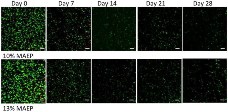
Figure 1.

Confocal fluorescence microscopy representative images of sections taken from the center of the hydrogels stained with Live/Dead reagent at various time points. Live cells fluoresce green and dead cells stain red. Autofluorescence of the hydrogels at the wavelengths used for the dead dye component was observed after day 7 and therefore the images for days 14-28 only show the live stain. Rat MSCs were encapsulated in hydrogels composed of 10 and 13 mol% MAEP and incubated in complete osteogenic medium until the desired time point. Scale bar = 100μm.
