Figure 10.

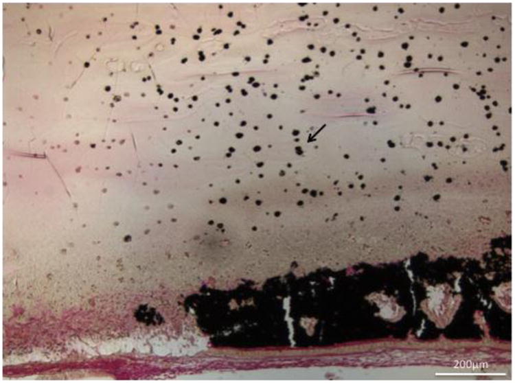
Histological section (von Kossa stain- mineralized tissue stains black) of cell induced mineralization within a 13 mol% MAEP cell-laden hydrogel (arrow) at the 4 week timepoint. Scale bar is 200μm.

Histological section (von Kossa stain- mineralized tissue stains black) of cell induced mineralization within a 13 mol% MAEP cell-laden hydrogel (arrow) at the 4 week timepoint. Scale bar is 200μm.