
Figure 5.
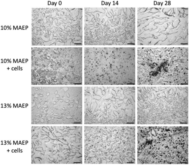
Representative histological sections (von Kossa stain- mineralized tissue stains black) of center-cut sections of hydrogels composed of 10 and 13 mol% MAEP with and without rat MSCs. Hydrogels were incubated in complete osteogenic medium until the desired time point. Scale bar is 100μm.