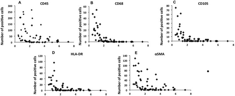
Fig. 6. Spatial distribution of CD45 (A), CD68 (B), CD105 (C), HLA-DR (D), and SMA (E) positive cells within the ILT.

Data from 6 patients is displayed. Two samples per patient were examined. Results demonstrate presence of cells expressing myeloid, mesenchymal and progenitor cell markers, which are largely located within the luminal region. Few cells of any type were observed within the medial or abluminal portions of the ILT.
