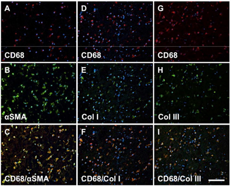
Fig. 7. Coexpression of fibrocyte markers in cells from the ILT.

Cells within the ILT express a unique “fibrocyte-like” phenotype. Cells colocalizing expression of CD68+/SMA+ (A-C), CD68+/Collagen I+ (D-F), and CD68+/Collagen III+ cells (G-I) were all observed. Red: CD68, Blue: DAPI, Green: SMA (B), Collagen I (E), Collagen III (H). Scale bar = 100 μm.
