
FIGURE 1. Effects of AdSAT1 transduction, cycloheximide, DFMO, putrescine, N1-acetylspermidine, α-MeSpd, Me2Spm and BENSpm on cell viability.
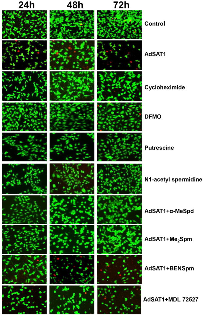
Viability of 293T cells was examined using a Live/dead cell imaging kit at 24, 48 and 72 h after seeding with no treatment (control), or AdSAT1 transduction, or treatment with cycloheximide (1 μg/ml), or with DFMO (5 mM), putrescine (5 mM) or N1-acetylspermidine (5 mM) or AdSAT1 transduction with added BENSpm (10 μM), α-MeSpd (100 μM), Me2Spm (100 μM). The concentrations of BENSpm, α-MeSpd and Me2Spm were chosen based on previous reports [9, 42]. Cells were stained with calcein and dead-red to visualize live and dead cells, respectively. Live cells fluoresce a bright green, whereas dead cells with damaged membranes fluoresce a red color. Representative images from three independent experiments are shown.