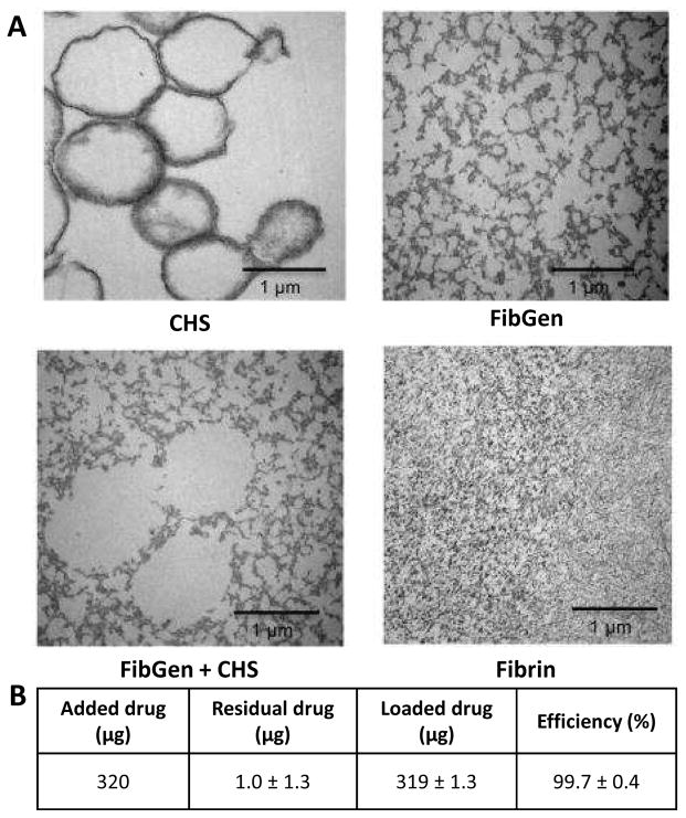
Figure 2. Characterization of collagen hollow sphere and FibGen morphology and drug release efficiency using transmission electron microscopy (TEM).
(A) Collagen hollow sphere morphology was observed using transmission electron microscopy. Sphere size and shape was maintained when CHS were mixed with FibGen. FibGen porosity was not altered with addition of CHS. Fibrin porosity was markedly smaller compared to FibGen. (B) Drug loading efficiency was assessed by loading initial 320 μg into CHS and measuring residual drug release (1.0±1.3μg) and loading efficiency was calculated (99.7%).

