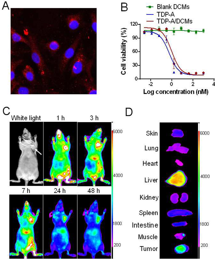
Figure 5.
(A) Confocal images showing the intracellular uptake of DiD fluorescently labeled DCMs in MDA-MB-231 cells after 4-h incubation. (B) The in vitro cytotoxicity of TDP-A/DCMs against MDA-MB-231 cells after 72 h incubation, measured by MTS assay. TDP-A loading in DCMs was 1 mg/mL. (C) In vivo NIRF optical images of orthotopic MDA-MB-231 tumor xenograft bearing mice after intravenous injection of DiD-TDP-A/DCMs. (D) Ex vivo NIRF image of dissected organs and tumor obtained at 48 h post-injection.

