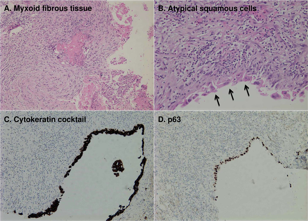
Figure 2. Pathologic report: Squamous Cell Carcinoma (at least in-situ).
Loose myxoid fibrous tissue with extensive inflammation, including lymphocytes, histiocytes, and eosinophils (A). Fragments are focally lined by atypical squamous cells, which form aggregates (arrows) (B). Immunohistochemical staining of the lining epithelial cells is positive for cytokeratin cocktail (C) and p63 (D). It could not be determined by pathologic appearance whether this squamous lesion represented a primary SCC or a metastatic SCC with subsequent cystic change.

