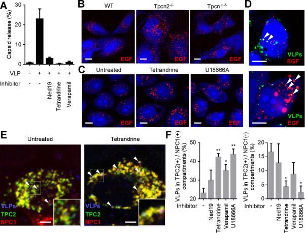
Fig. 3. Blocking TPC function impacts EBOV entry through endosomal compartments.
(A) VLPs loaded with β-lactamase were used to measure membrane fusion and virus capsid release into the cytoplasm after each treatment (as for fig. S10). The number of cells showing signal were divided by the number of total cells. (B) Evaluation of EGF trafficking in TPC knockout cells. Representative confocal images of WT, Tpcn2−/− and Tpcn1−/− MEFs incubated with AlexaFluor555-EGF. (C) Evaluation of EGF trafficking in tetrandrine or U18666A-treated cells. Representative confocal images of HeLa cells incubated with AlexaFluor555-EGF (red) in the presence or absence of tetrandrine or U18666A. (D) Colocalization of Ebola VLPs and EGF. HeLa cells were incubated with AlexaFluor555-EGF (red) for 30 min followed by Ebola VLPs (green) for 3.5 h in the presence of tetrandrine. VLPs were stained with a GP-specific antibody. Examples of colocalized particles are indicated by arrowheads. Scale bars, 10 μm for (B), (C) and (D). (E) Effect of tetrandrine on colocalization of Ebola VLPs with TPC2- and/or NPC1-positive endosomes was measured. HeLa cells overexpressing GFP-tagged TPC2 (green) and myc-tagged NPC1 (red) were pretreated with inhibitors and incubated with VLPs (blue) for 4 h. Insets show magnified areas of the image and arrowheads indicate examples of VLPs that are associated with the TPC2(+)/NPC1(−) compartment (left panel) or the TPC2(+)/NPC1(+) compartment (right panel). Scale bars, 5 μm. (F) In the presence of the indicated inhibitors, the ratio of VLPs colocalizing with the TPC2(+)/NPC1(+) compartment (left) or the TPC2 (+)/NPC1(−) compartment (right) was calculated. * and ** indicate P < 0.05 and P < 0.005, respectively, using unpaired Student's t-test to compare treated to untreated cells. Data are mean ± SEM (n = 3 or 4).

