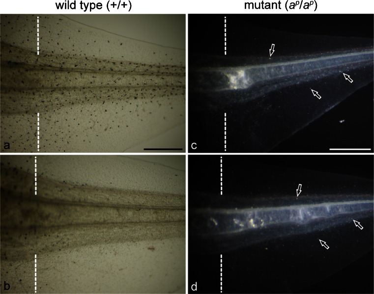
Fig. 1.
Expression of pigment cells in the 6-day regenerating tail in the absence (a, c) or presence (b, d) of phenylthiourea (PTU); amputated at stage 50. a, b Wild type regenerating tail observed under transmitted light. c, d Mutant regenerating tail observed under incident light. Dashed lines indicate amputation level. Many melanophores appeared in the wild type regenerating tail in the absence of PTU (a). However, few melanophores appeared in the wild type regenerating tail in the presence of PTU (b). In contrast, white pigment cells (arrows) appeared in the mutant regenerating tail in both the absence (c) and presence (d) of PTU. Bar 500 μm

