Fig. 5.
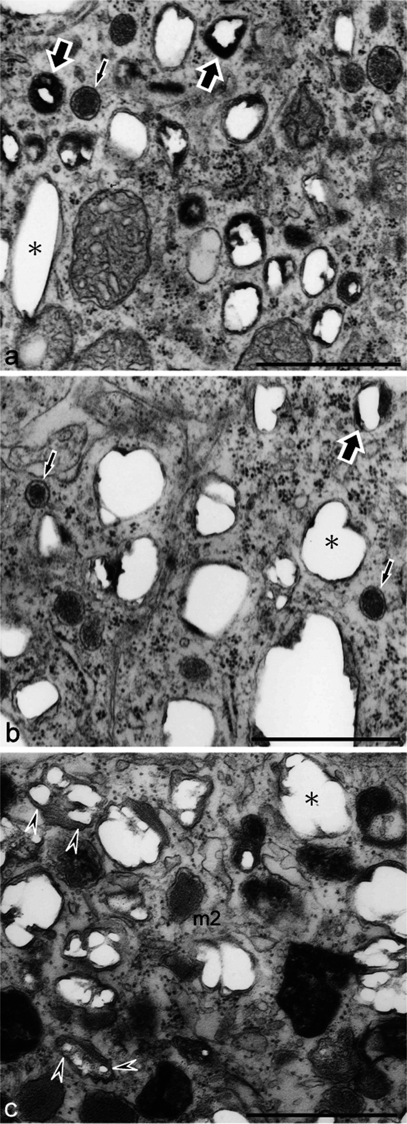
Reflecting platelet organellogenesis in iridophores and white pigment cells. a, b Ultrastructure of wild type (a) and mutant (b) iridophores that were allowed to differentiate in culture. c Ultrastructure of white pigment cells in the mutant regenerating tail. Spherical vesicles with electron-dense material (a, b, small arrows) were present in both wild type and mutant iridophores. Spherical vesicles subsequently accumulated crystals that were lost partially during fixation and thin-sectioning, leaving “partial holes” (large arrows) in the sections. Mature reflecting platelets were characterized by “empty holes” (asterisk). Reflecting platelets of wild type iridophores were rectangular (a, asterisk); however, those of mutant iridophores were irregular in size and shape (b, asterisk). White pigment cells in the mutant contained irregular-shaped reflecting platelets (c, asterisk) and stage II melanosomes with internal lamellar structures (m2). Note that reflecting platelets in white pigment cells were formed from stage II melanosomes (c, arrowheads), but not from spherical vesicles that were observed in iridophores (b). Bar 1 μm

