Fig. 6.
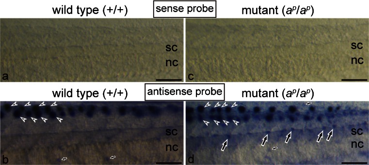
Spatial expression of the ferritin H subunit mRNA in the middle region of the tail in the wild type (a, b) and the mutant (c, d) at stage 48. Whole mount in situ hybridization (WISH) was performed by using sense (a, c) or antisense (b, d) digoxigenin (DIG)-labeled RNA probes. Tadpoles were bleached to remove melanin before hybridization in this experiment. With a sense probe of the ferritin H subunit mRNA, no staining was observed in the tail of both the wild type (a) and the mutant (c) in the negative control. Use of an antisense probe in WISH detected strong staining in the lateral lines (arrowheads) of both the wild type (b) and the mutant (d). In addition, specific expression of the ferritin H subunit mRNA was detected in white pigment cells (d, large arrows), which were present around the dorsal side of the spinal cord (sc) in the mutant (nc notochord). Although melanophores were present around the dorsal side of the spinal cord in the wild type, no staining was observed in melanophores (b). Note that staining was also detected in some epidermal cells (small arrows) in both the wild type (b) and the mutant (d). Bar 100 μm

