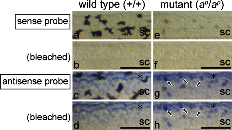
Fig. 7.
Expression of the ferritin H subunit mRNA in white pigment cells but not in melanophores. Photographs of the posterior region of the wild type tail (a-d) and the mutant tail (e-h) at stage 48. WISH was performed with sense (a, b, e, f) or antisense (c, d, g, h) DIG-labeled RNA probes (sc spinal cord). Tadpoles were bleached after BM purple staining. The same fields before (a, c, e, g) and after (b, d, f, h) bleaching are shown. In the negative control, with a sense probe of the ferritin H subunit mRNA, no staining was observed in the tail of the wild type (a, b) or the mutant (e, f). Before bleaching, dendritic black melanophores (a) were distinguished from punctate white pigment cells (e), which appeared brown under transmitted light. Melanin was bleached effectively in both melanophores (b) and white pigment cells (f). Staining with an antisense probe indicated that white pigment cells (g, h, arrows), but not melanophores (c, d), expressed the ferritin H subunit mRNA. Note that staining was also detected in the dorsal longitudinal anastomosing vessel (asterisks) in both the wild type (c, d) and the mutant (g, h). Bar 100 μm

