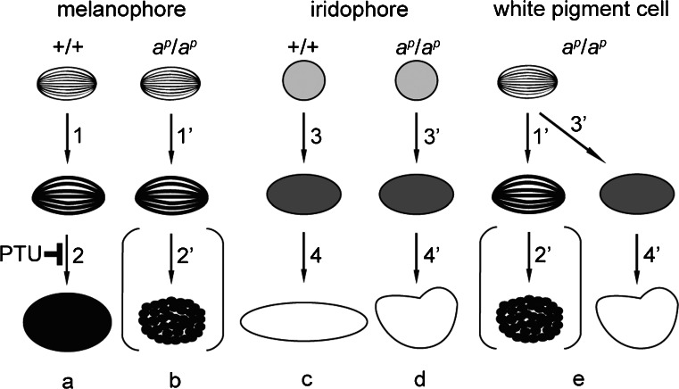
Fig. 9.
Representation of pigment organellogenesis in melanophores (a, b), iridophores (c, d), and white pigment cells (e). a, b Melanosome formation in wild type (a) and mutant (b) melanophores. c, d Reflecting platelet formation in wild type (c) and mutant (d) iridophores. e Pigment organellogenesis in white pigment cells in the mutant. In wild type melanophores, melanin deposition occurs in stage II melanosomes to form partially melanized stage III melanosomes (a, 1) and then fully melanized stage IV melanosomes are formed (a, 2). Melanin is also deposited in stage II melanosomes to form stage III melanosomes (b, 1’) in mutant melanophores; however, few melanosomes become fully melanized (b, 2’). PTU inhibits melanosome maturation from stage III melanosomes to stage IV melanosomes in melanophores. In wild type iridophores, spherical vesicles with electron-dense material accumulate crystals (c, 3), which grow larger and exhibit rectangular reflecting platelets (c, 4). Crystals are also accumulated in spherical vesicles with electron-dense material in mutant iridophores (d, 3’); however, reflecting platelets become irregular in shape (d, 4’). In white pigment cells, melanosome formation (e, 1’, 2’) occurs in the same manner as described in mutant melanophores (b, 1’, 2’). In addition, some stage II melanosomes accumulate crystals in white pigment cells (e, 3’), and irregular-shaped reflecting platelets are formed (e, 4’)

