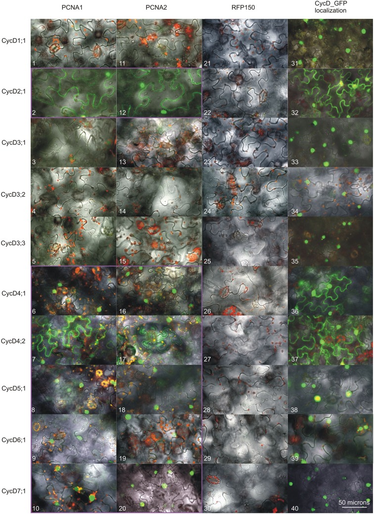
Figure 2.
Analysis of D-type cyclins subcellular localization and the formation of complexes with either PCNA1 or PCNA2. Confocal images of N. benthamiana leaf cells expressing transiently analyzed open reading frames. Split GFP complex formed between PCNA1_CtermGFP and (1) CycD1;1_NtermGFP, (2) CycD2;1_NtermGFP, (3) CycD3;1_NtermGFP, (4) CycD3;2_NtermGFP, (5) CycD3;3_NtermGFP, (6) CycD4;1_NtermGFP, (7) CycD4;2_NtermGFP, (8) CycD5;1_NtermGFP, (9) CycD6;1_NtermGFP, (10) CycD7;1_NtermGFP. Split GFP complex formed between PCNA2_CtermGFP and (11) CycD1;1_NtermGFP, (12) CycD2;1_NtermGFP, (13) CycD3;1_NtermGFP, (14) CycD3;2_NtermGFP, (15) CycD3;3_NtermGFP, (16) CycD4;1_NtermGFP, (17) CycD4;2_NtermGFP, (18) CycD5;1_NtermGFP, (19) CycD6;1_NtermGFP, (20) CycD7;1_NtermGFP. Split GFP complex formed between RFP150_CtermGFP and (21) CycD1;1_NtermGFP, (22) CycD2;1_NtermGFP, (23) CycD3;1_NtermGFP, (24) CycD3;2_NtermGFP, (25) CycD3;3_NtermGFP, (26) CycD4;1_NtermGFP, (27) CycD4;2_NtermGFP, (28) CycD5;1_NtermGFP, (29) CycD6;1_NtermGFP, (30) CycD7;1_NtermGFP. (31) CycD1;1_GFP, (32) CycD2;1_GFP, (33) CycD3;1_GFP, (34) CycD3;2_GFP, (35) CycD3;3_GFP, (36) CycD4;1_GFP, (37) CycD4;2_GFP, (38) CycD5;1_GFP, (39) CycD6;1_GFP, (40) CycD7;1_GFP. All the images are overlays of the bright field, autofluorescence of chlorophyll (red) and GFP fluorescence (green). The PCNA/CycD complexes are in the magenta frame. This result is representative of three independently repeated experiments.

