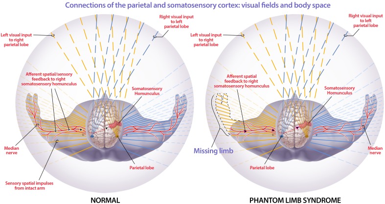
Figure 5.
Connections of the parietal and somatosensory cortex: visual fields and body space. The orange lines are visual and non-visual information sent to the right parietal cortex while the blue lines are visual and non-visual information sent to the left parietal cortex. The red sensorimotor cortices are depicted in each hemisphere and the corresponding red lines illustrate the processed sensorimotor information that is filled in within 3D default space. The figure depicts how the right parietal lobe and sensory motor cortex, in a healthy individual, receives visual information from the left eye, and sensory information from the left side of the body. The parietal lobe spatially maps this information so that the mind can spatially locate stimuli. In phantom limb syndrome, when the left arm has been amputated, visual information that the limb is gone is received by the left eye and processed so that the visual information filled into the 3D default space does not include a left arm within the visual space. Though there is no longer sensorimotor information flowing from the now amputated limb to the cortex, the sensorimotor and other networks in the brain are still intact. This leads to intrinsically-generated sensations that are integrated within the 3D default space, generating an intact non-visual representation of the limb and phantom limb sensations. Phantom limb pain is likely due to the integration of this contradictory sensory information and subsequent representation within 3D default space. The solid lines extending through the body illustrate the information, from the corresponding cortex, that is filled into the 3D default space. (Figure and figure description reproduced with permission from copyright holder. Figure by Michael Jensen MSMI, CMI).

