
Figure 2.
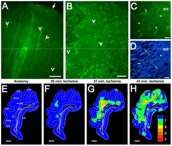
Diffuse WMI is associated with markedly less cerebral gray matter injury in preterm human autopsy cases (A-D) and in a pre-clinical large animal model of WMI in preterm fetal sheep (E-H). From a preterm human autopsy case diagnosed with early diffuse WMI, the relative amounts of cell death are compared in parietal cerebral cortex (A) relative to a diffuse deep cerebral white matter lesion (B). From these images of fluorescently-labeled degenerating cells (arrowheads provide typical examples), the paucity of degenerating cells in the cortex (A) relative to the white matter (B) can be appreciated. The large arrow in A indicates the pial surface. (C) This image shows an early small focal deep white matter lesion (arrows), adjacent to the subventricular zone (SVZ), that was less than a millimeter in diameter. Such lesions typically evolve to microscopic necrosis. (D) This image visualizes all of the cells present in the region shown in C and demonstrates the high density of cells in the SVZ relative to the adjacent deep white matter in the center of the image. (E) Anatomical diagram of the preterm fetal sheep brain at 0.65 gestation, which is approximately equivalent to the preterm human brain at 26-28 weeks gestation. Animals were analyzed for the distribution of degenerating cells that occurred in response to cerebral hypoxia-ischemia of durations of 30 minutes (min.) (F) 37 minutes (G) or 45 minutes (H). The pseudocolor maps in F-H indicate the overlapping distributions of degenerating cells observed in four animals at the level of parietal cortex. The pseudocolor probability scale (1, low; 7, high) indicates the relative frequency with which cell death was observed throughout this level of the brain. Note that WMI in the periventricular white matter (PVWM), superficial white matter (SWM), internal capsule (IC) and corpus callosum (CC) increased as the duration of ischemia was increased from 30 to 45 min. Injury to the cerebral cortex (CTX) and hippocampus (HIP) was rarely observed until the duration of ischemia was very prolonged to 45 min., which also coincided with a pronounced increase in necrotic WMI (H). Panels A-D adapted from Back et al., 2005.32 Panels E-H adapted from Riddle et al., 2006.34 Scale Bars: = 200 μm (A); 100 μm (B-D); 2 mm (E-H).