. 2015 Jul 30;4:e07607. doi: 10.7554/eLife.07607

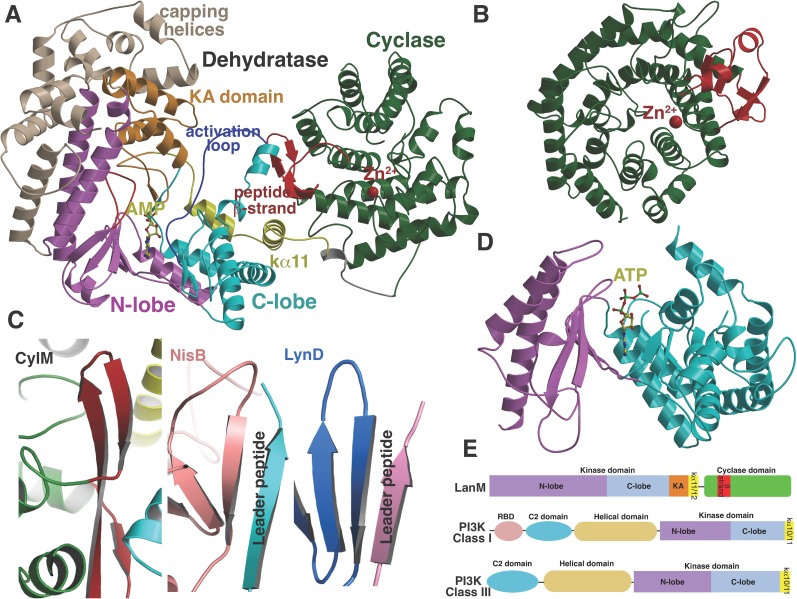
Figure 2. (A) Overall structure of CylM.

(B) Structure of the class I lanthipeptide cyclase NisC illustrating the structural homology with the C-terminus of CylM. (C) Comparison of the putative peptide-binding β-strands of CylM with the peptide binding regions of other RiPP biosynthetic enzymes including NisB (involved in nisin biosynthesis, PDB 4WD9) and LynD (involved in cyanobactin biosynthesis; PDB 4V1T). (D) Structure of the lipid kinase PI3K that shares homology with the dehydration domain of CylM. (E) Domain organization of LanMs in comparison with that of lipid kinases. RBD, Ras-binding domain.

DOI: http://dx.doi.org/10.7554/eLife.07607.006

Figure 2.

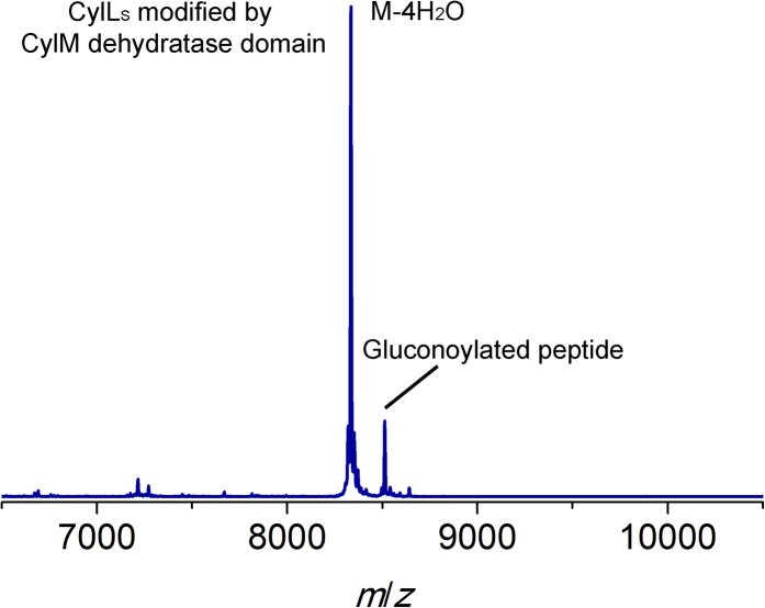
Figure 2—figure supplement 1. MALDI-TOF mass spectrum for CylLS modified by the CylM dehydratase domain in Escherichia coli.

Figure 2—figure supplement 1.

Calculated M—4 H2O: 8330, average mass; observed M—4 H2O + H+: 8334, average mass. Partial gluconoylation at the N-terminus of dehydrated CylLS occurred when expressing the peptide in E. coli BL21(DE3), resulting in a +178 Da peak in addition to the desired peptide mass (Aon et al., 2008).
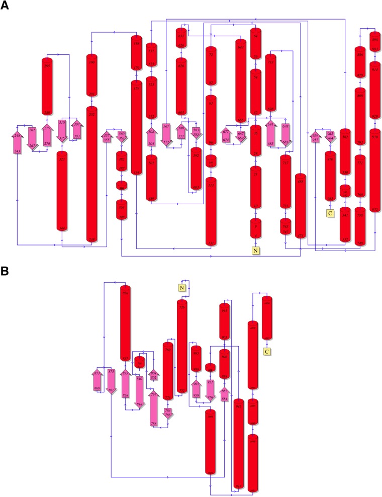
Figure 2—figure supplement 2. Topology diagrams for (A) CylM and (B) PI3 kinase P110γ.

Figure 2—figure supplement 2.

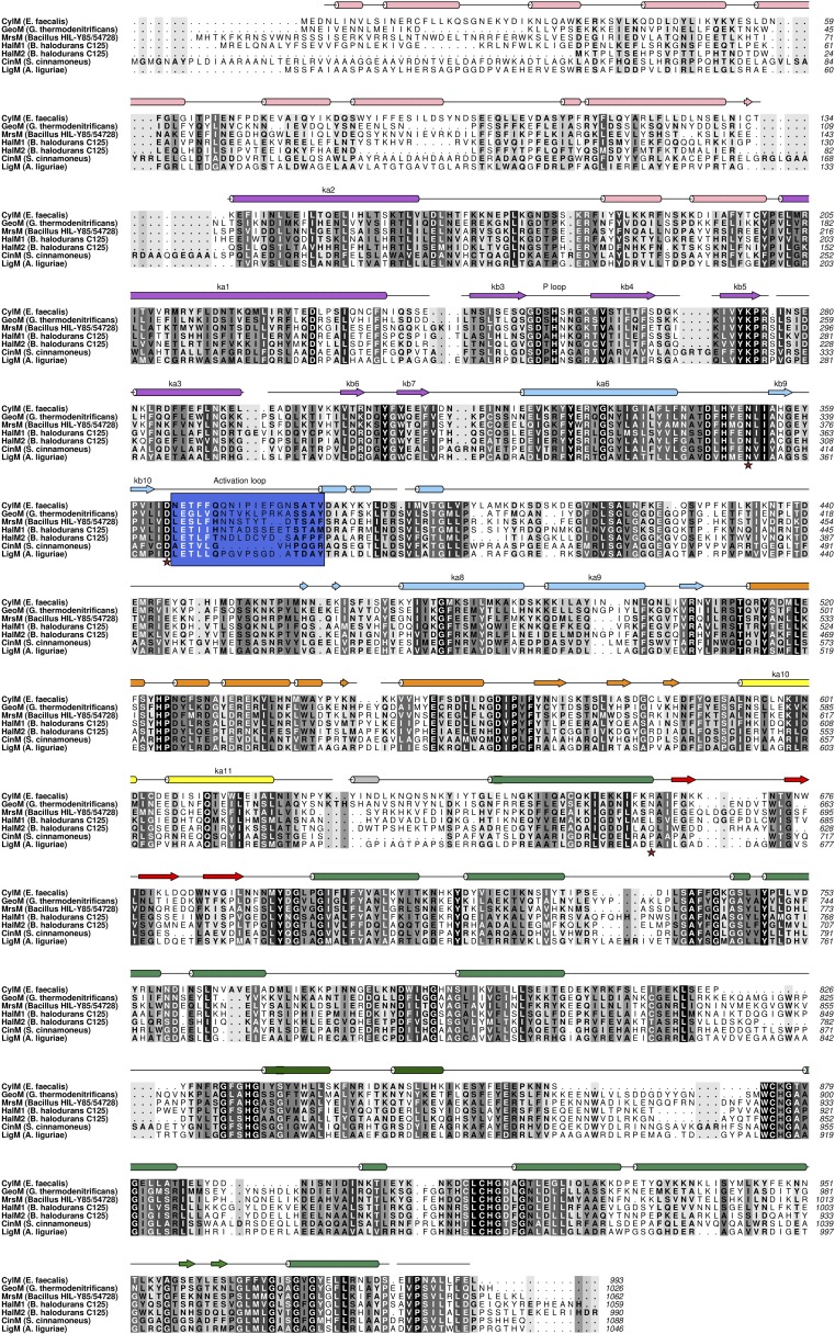
Figure 2—figure supplement 3. Structure based alignment of biochemically characterized LanM enzymes.

Figure 2—figure supplement 3.

Secondary structural elements are colored as in Figures 2A, 3A.