Figure 6.
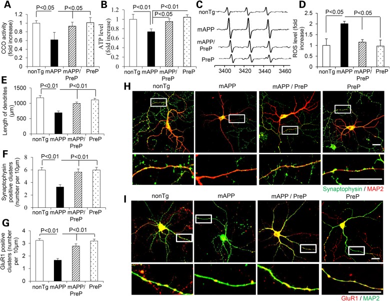
Effect of neuronal PreP on mitochondrial function, ROS and synaptic density in cultured AD neurons. Complex IV/CCO activity (A), ATP levels (B) and cellular ROS (C and D) were measured in hippocampal neurons derived from the indicated Tg mice. Representative EPR spectra (C) and quantification (D) for ROS signals in the indicated groups of cells. Data are expressed as fold-increase relative to nonTg neurons. (E–I) Effect of PreP on length and synaptic density of neuronal dendrites. Hippocampal culture neurons from the indicated Tg mice in vitro day 12 were stained with MAP2 (a marker for dendrites, H and I) and Syn (pre-synaptic marker, green, H) or GluR1 (post-synaptic marker, red, I). Quantification of a total length of dendrites (E), number of synaptophysin-positive clusters (F) and GluR1-positive clusters (G) per micron of dendrite length was performed in the indicated groups of cells. (H) The representative staining images for synaptophysin (green) and MAP2 (red), or for (I) GluR1 (red) and MAP2 (green) in the indicated groups of cells. The lower panels of H and I are enlarged images corresponding to above framed images.

