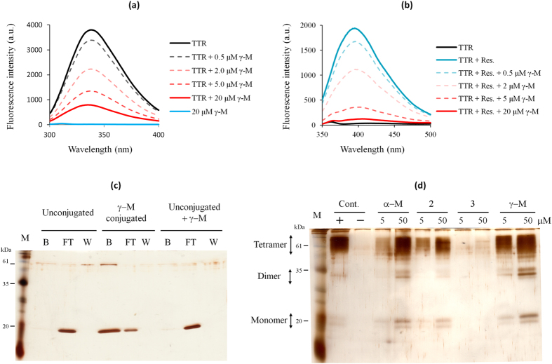
Figure 2. γ-M binds to the T4-binding sites and stabilizes the V30M tetramer.
(a) Intrinsic fluorescence spectra (300–400 nm) of V30M and upon the addition of γ-M by excitation at 280 nm. (b) The fluorescence spectra (350–500 nm) of resveratrol (5 μM) bound to V30M and upon the addition of γ-M by excitation at 320 nm. (c) Pull-down assay using CN-Br activated Sepharose. V30M was incubated with γ-M conjugated to Sepharose or Sepharose alone. B indicates the bead fractions, FT indicates the flow-though fractions and W indicates the final wash fractions. V30M was observed in the lane for the γ-M conjugated beads, but not in that for Sepharose alone. (d) Effect of γ-M on the acid-mediated quaternary structural changes of TTR (10 μM). The + lane indicates the positive control incubated at pH 8.0 without the compounds. The - lane indicates the negative control incubated at pH 4.5 without the compounds.

