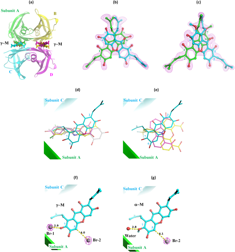
Figure 3. Crystal structures of V30M in complex with γ-M and α-M.
(a) Overall structure of V30M in complex with γ-M. (b) Omit Fourier map of γ-M bound to TTR contoured at 2.8 σ. (c) Omit difference Fourier map of α-M bound to TTR contoured at 3.3 σ. (d) A comparison of the binding directions with the known stabilizers. The carbon atoms of tafamidis (PDB ID: 3TCT), resveratrol (1DVS) and glabridin (4N87) are colored yellow, magenta and silver, respectively. (e) A comparison of the binding directions with the known stabilizers with a tricyclic ring system. The carbon atoms of PHENOX (1DVY) and dibenzofuran-4,6-dicarboxylic acid (1DVU) are colored magenta and yellow, respectively. Anomalous difference Fourier maps of Br derivatives of the γ-M (f) and α-M (g) complexes contoured at 5.0 σ are also shown.

