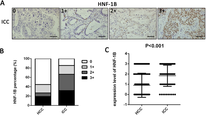
Figure 2. HNF-1B expression is associated with different pathologic types of primary tumor.
(A) HNF-1B immunohistochemical staining patterns in ICC tissues. Representative sections showing HNF-1B expression in 0, 1+, 2+ and 3 + (sacle bar = 100 μm). (B) The differences of HNF-1B expression percentage between HCC patients and ICC patients. (C) The significant differences of HNF-1B expression level between HCC patients and ICC patients (Mean±SD).

