Fig. 4.
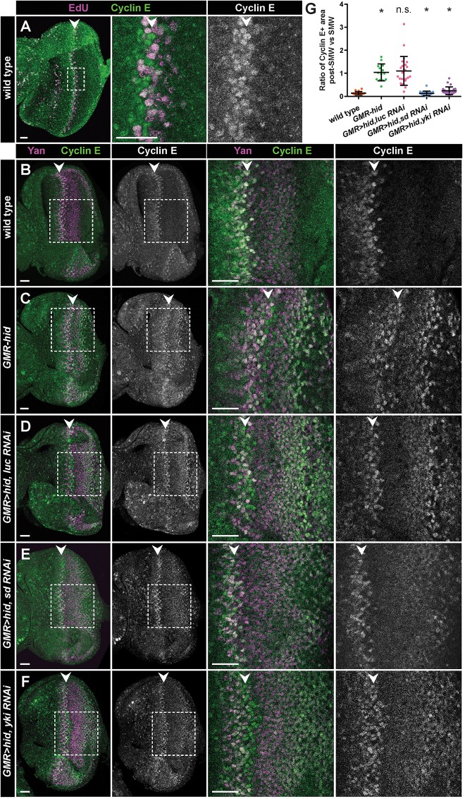
Sd and Yki are required for elevated Cyclin E levels during CP. (A) Eye disc labeled with EdU (magenta) and anti-Cyclin E (green) antibodies. Cyclin E accumulates prior to and during S phase of the SMW (arrowhead). Box in left panel indicates area of magnification in middle and right panels. (B-F) Eye discs of the indicated genotypes stained with anti-Cyclin E (green) and anti-Yan (marker of undifferentiated cells; magenta) antibodies. Boxes indicate areas shown at higher magnification on the right. Arrowheads indicate SMW. (G) Quantification of Cyclin E staining in Yan+ cells of the indicated genotypes. The ratio of post-SMW Cyclin E staining versus SMW Cyclin E staining is displayed (see supplementary Methods for details). Each circle represents the ratio calculated for a single disc, and bars represent mean and one standard deviation. For each genotype, n≥14 discs. *P≤1.8×10⁻⁷. Significance was calculated for wild type (w1118) versus GMR-hid, GMR-hid versus GMR>hid, luc RNAi, GMR>hid, luc RNAi versus GMR>hid, sd RNAi, and GMR>hid, luc RNAi versus GMR>hid, yki RNAi. n.s., not significant. Anterior is oriented to the left. Scale bars: 20 μm.

