Fig. 6.

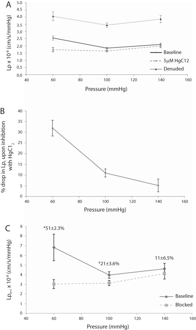
Lp of the rat aorta ex vivo. Figure 1 shows the experimental setup. A: Lp(ΔP) for an excised aorta from 300- to 350-g male SD rats, first without (bold solid) and then with (dashed) the AQP blocker HgCl2, and then after endothelial denudation (thin solid), with all nine Lp values taken on each vessel. HgCl2 reduced every vessel's Lp significantly at every pressure. B: replot of these data as means ± SE of the percent drop in Lpt with HgCl2 versus ΔP. HgCl2 reduced Lpt (32 ± 4%) at 60 mmHg and less [yet significantly different (P = 0.000026) and nonzero] at 100 (11 ± 2%) and 140 mmHg (5 ± 3%). C: since HgCl2 does not change Lpm + I (Table 5), changes in Lpt reflect changes in endothelium + SI Lpe + i (points); numbers above the points are means ± SE of percent drops in Lpe + i with HgCl2. These differences are distinguishable (P < 0.005) for 60 mmHg versus the other pressures only. (This figure and these statistics neglect the lone outlier that showed nearly no difference between Lpt and Lpm + I, and thus a huge Lpe + i, at 60mmHg only; the mean ± SE for the percent drop including this outlier at 60 mmHg was 56 ± 7.4%). Lpe + i dropped by more than half at 60 mmHg. n = 6.
