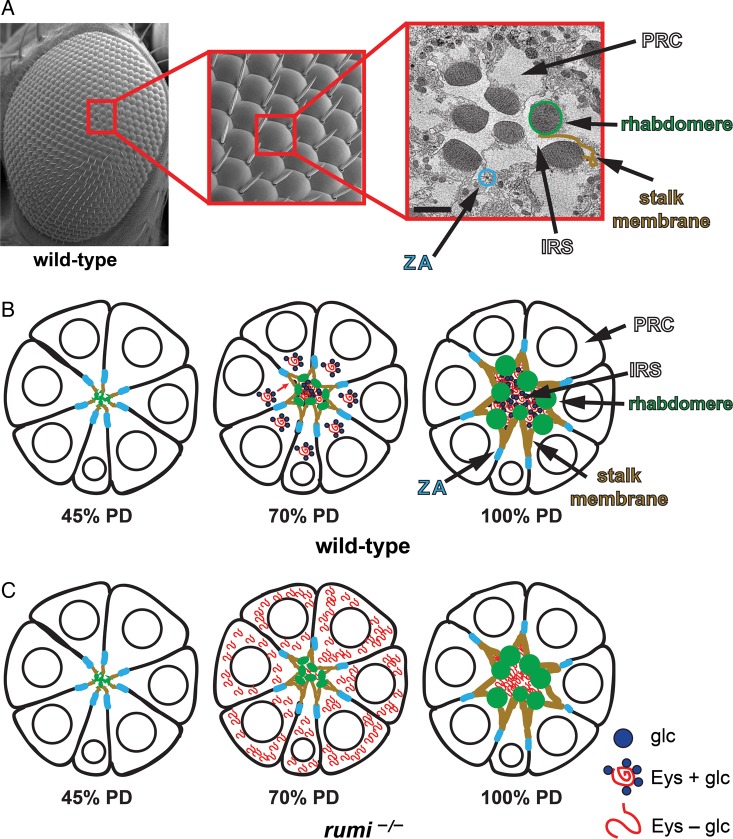
Fig. 3.
A model for the regulation of Eyes shut and IRS formation by Rumi. (A) A scanning electron micrograph (SEM) of the adult fly eye with its 760–800 ommatidia is shown to the left. The close-up shows a number of ommatidia, with sensory bristles decorating alternating corners of each ommatidium. To the right is a transmission electron micrograph of a single ommatidium, showing seven photoreceptor cells (PRCs). SEM images are courtesy of Jessica Leonardi. (B) Schematic drawings of a developing ommatidium. At 45% PD, the apical surfaces of photoreceptors contact one another and rhabdomere formation has not started. By 70% PD, disc-shaped rhabdomeres are formed at the apical side of photoreceptors, and secretion of Eyes shut (Eys) has separated the apical surfaces of the rhabdomeres and has formed a continuous IRS. Most of the Eys protein, which is O-glucosylated by Rumi in wild-type animals, is found in the IRS. By 100% PD, rhabdomeres have assumed their round adult morphology and are well separated by an Eys-filled IRS. (C) In rumi null eyes, a significant amount of Eys, which should lack O-glucose and is likely misfolded, remains in the PRC body. At this stage, only a fraction of Eys is secreted into the IRS, which is smaller than the wild-type IRS at the same developmental time. By 100% PD, rumi mutants do not show Eys accumulation in the PRC bodies anymore and accumulate Eys in the IRS, whose size is comparable with wild-type IRS at this stage. However, the rhabdomere attachments are not resolved, and the IRS is not continuous, suggesting that rhabdomere separation needs to occur in a critical time window during development. The ommatidium schematics are adapted from Knust (2007). Circles in PRCs depict the nuclei. Glc, glucose; ZA, zonula adherens. This figure is available in black and white in print and in color at Glycobiology online.

