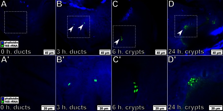
FIG 3.
Tracking the bacterial position during initiation of colonization. For all images, bacteria were labeled with probes specific for 16S rRNA (green), and squid tissue was stained with Alex Fluor 633-phalloidin (blue). (A′ to D′) Enlargements of the boxed regions in panels A to D, respectively. Magnifications, ×40. (A, A′) Before exposure, no bacteria were visible within the light organ of the host. (B, B′) After 3 h of exposure, bacteria (arrowheads) have associated with the host and begin to be visible within the light organ ducts, located immediately in the interior of the pores (Fig. 1C). (C, C′) By 6 h postexposure, bacteria have migrated into the light organ and have begun to colonize the crypt space. (D, D′) After 24 h of exposure, the host is bioluminescent and the symbionts are visible throughout the crypts.

