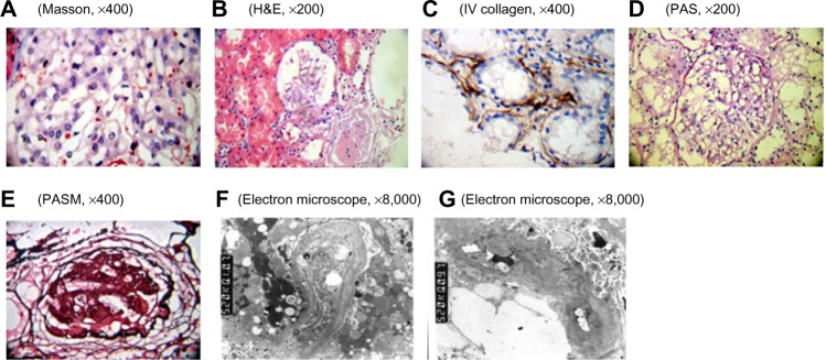
Figure 1.

Pathological examination of the kidney by using light microscopy and electron microscopy.
Notes: (A) mesangial mild proliferation; (B) mesangial mild proliferation and glomerulosclerosis; (C) renal tubular atrophy and renal tubular basement membrane type IV collagen deposition; (D) GBM thickening; (E) glomerular sclerosis; (F and G) irregular thickening, thinning, and splitting of GBM. Electron-dense material deposited in the membrane. Mesangial matrix severely increased. There were dense deposits showing cord-like distribution. Part of tubular cell vacuolar degeneration and tubular basement membrane segmental thickening.
Abbreviations: GBM, glomerular basement membrane; H&E, hematoxylin & eosin; PAS, periodic acid–Schiff; PASM, periodic acid-silver motheramine.
