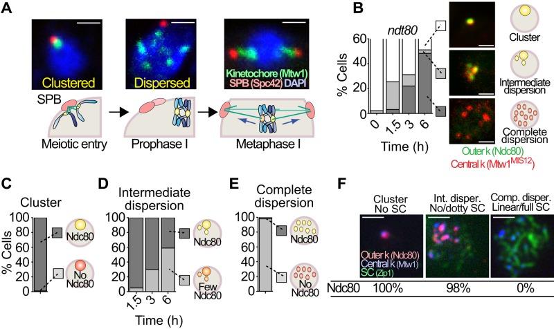
FIGURE 1:
Centromere repositioning and outer kinetochore disassembly. (A) Representative pictures of diploid cells carrying markers for the kinetochore (MTW1-GFP) and the SPBs (SPC42-DsRed) progressing into meiosis. The cells enter meiosis with clustered centromeres, and centromeres disperse in prophase and then reattach, with sister chromatids attaching to microtubules from the same pole and homologues drawn to opposite poles. (B) A diploid strain with marked central and outer kinetochore proteins (MTW1-3xmCherry and NDC80-GFP) was released into meiosis, and cells with clustered centromeres and Ndc80 present, intermediate dispersion (<4 Mtw1 foci), or complete dispersion (>4 Mtw1 foci) and no Ndc80 were scored (n ≥ 100). Representative cells. Scale bar, 1 μm. (C) From B, the cells with clustered centromeres with (dark gray) or without Ndc80 (light gray) were scored. (D) From B, cells with intermediate dispersion and strong (dark gray) or weak (light gray) signals for Ndc80 were scored. (E) From B, cells with full dispersion and with (dark gray) or without Ndc80 (light gray) were scored. (F) In a diploid strain carrying the kinetochore markers MTW1-3xmCherry and NDC80-GFP, cells at different stages of synaptonemal complex (Zip1) assembly (none, dotty, or linear/full) were scored for the presence of Ndc80. Scale bar, 1 μm.

