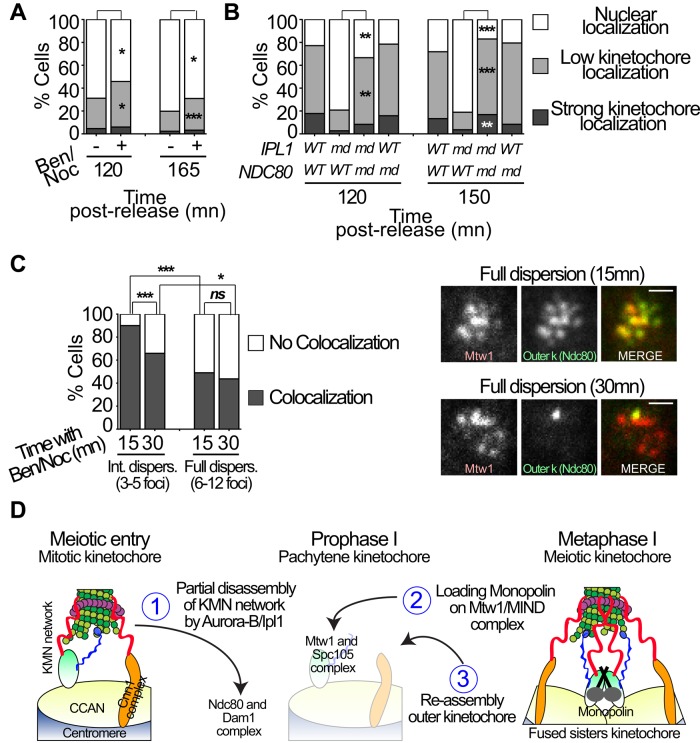
FIGURE 7:
Release of kinetochore–microtubule associations and shedding of outer kinetochores allow monopolin loading. (A) Monopolin loading on kinetochores with or without release of kinetochore–microtubule attachments. ipl1-md diploid cells were sporulated and released from a pachytene arrest (PGAL1-NDT80 GAL4-ER) at 6 h after the induction of meiosis by the addition of 5 μM β-estradiol (T = 0 h). Microtubules were destabilized by the addition of benomyl (30 μg/ml) and nocodazole (15 μg/ml), which were added at the time of release from the pachytene arrest (protocol is summarized in Figure 6A). At 120 and 165 min after release from pachytene arrest, cells were categorized according to their Mam1-GFP localization (nuclear, low kinetochore localization, strong kinetochore colocalization; see Supplemental Figure 5 for representative images). n ≥ 83 cells for each time point. (B) Monopolin loading with and without the Ndc80 complex on kinetochores. Wild-type, ipl1-md, ndc80-md, or ipl1-md ndc80-md diploid strains were induced to enter meiosis and then released from pachytene by the addition of 5 μM β-estradiol at 4.5 h after the induction of meiosis. Cells were scored as in A. n ≥ 59 cells for each time point. (C) ipl1-md diploid cells expressing Ndc80-GFP and Mtw1-3xmCherry were switched to sporulation medium. The strains used were ndt80 mutants that arrest in pachytene. Benomyl (30 μg/ml) and nocodazole (15 μg/ml) were added to the cells 6 h after induction of meiosis (T = 0 h). The colocalization of Ndc80-GFP with individual Mtw1-mCherry foci was scored in cells with 3–5 dispersed kinetochores (intermediate dispersion) or those with 6–12 kinetochores (full dispersion). Colocalization is indicated by dark gray, and Mtw1-mCherry foci with no colocalizing Ndc80-GFP signal are indicated in white. Representative cells are shown. Scale bars, 1 μm. n ≥ 41 Mtw1 foci for each time point. Fisher's exact tests were used to evaluate the significance of observed differences in A–C. *p < 0.05, **p < 0.01, ***p < 0.001. (D) A model representing the steps of kinetochore remodeling in meiosis I.

