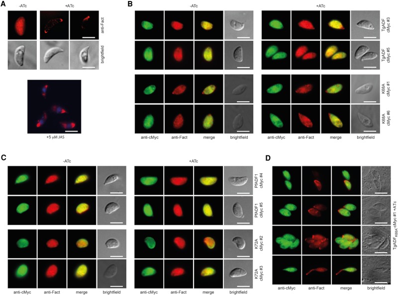
FIGURE 3:
Filamentous actin is distributed normally in the majority of complementing lines. (A) Parasites were grown with or without ATc for 68–72 h, and freshly released tachyzoites were fixed for immunofluorescence analysis. Suppression of TgADF-HA in the cKO line leads to an accumulation of actin at either (or both) of the two poles of the tachyzoite, which is similar to what is seen when tachyzoites are treated with JAS, a compound that stabilizes dynamic actin into filaments. (B, C) The majority of wild-type TgADF, mutant TgADFK68A, wild-type PfADF1, and PfADF1K72A-complementing clones display an even localization of F-actin throughout the cell in the absence and presence of ATc that is indistinguishable from what is observed in wild-type parasites. (D) A skewed distribution of actin is found in a small proportion (≤5%) of intracellular TgADFK68A-cMyc–expressing parasites, suggesting a polar accumulation of F-actin (as with JAS). Only very few parasites display an apical protrusion of actin (D, bottom). Scale bar, 5 μm.

