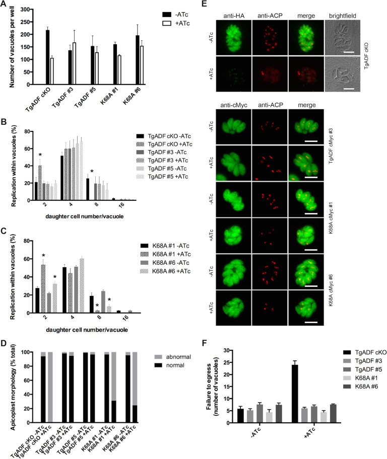
FIGURE 5:
Daughter cell formation and apicoplast segregation are impaired in TgADFK68A-cMyc–expressing parasites. (A) ATc-treated TgADF mutant parasite lines are slightly impaired in host cell invasion. Vacuoles were counted ∼24 h postinvasion and were visualized by using GAP45 antibodies by immunofluorescence analysis (IFA). Average of three independent experiments (± range). (B, C) TgADFK68A mutant parasites grown in ATc display a replication defect like that of the parent knockdown line (in which an increase in low vs. high nuclei number can be observed in the presence of ATc; asterisks). Daughter cells per vacuole were counted for each line and clone from the invasion experiments shown and normalized to the total vacuole number per condition (n = on average 300 vacuoles, ± range shown). (D, E) In parallel to the IFA carried out for invasion analysis (24 h postinvasion), IFA using anti-ACP antibodies was performed to investigate apicoplast segregation. Segregation of the apicoplasts as determined by distribution and number of plastid foci of fluorescence compared with no ATc (TgADF cKO) was greatly impaired in most TgADFK68A-cMyc–expressing parasites (clones #1 and #6 shown). Scale bar, 5 μm (E). (F) Egress (as determined by number of vacuoles in replicate assays) was not affected by mutation of the critical lysine residue. Average of five independent image panels from a representative egress assay.

