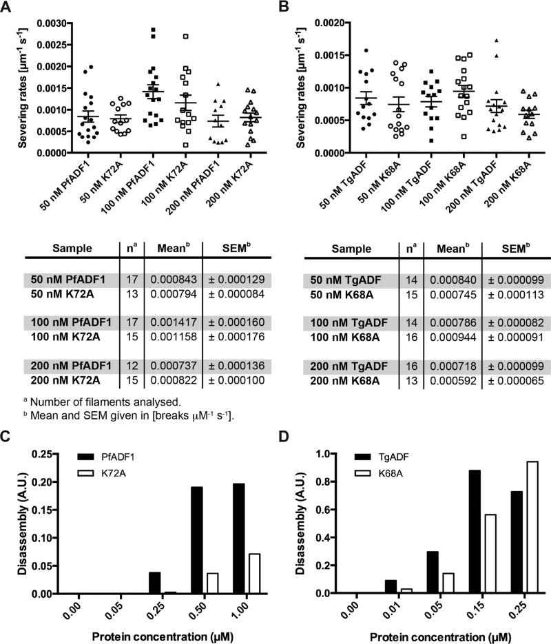
FIGURE 6:
TgADF is more efficient in disassembling F-actin than PfADF1. (A, B) TIRF microscopy analysis of cofilin-mediated F-actin severing by wild-type and mutant ADF proteins. Filaments were assembled from 1 μM G-actin (10% Oregon green–labeled, 0.1% biotinylated) in flow chambers and attached to biotin-PEG (0.1%)–coated glass slides by streptavidin. Plots of the normalized severing rates (break μm-1 s-1) in the presence of different protein concentrations show the individual data points, as well as the mean ± SEM. (C, D) F-actin disassembly assays using pyrene-labeled actin, induced by vitamin D–binding protein. A range of wild-type and mutant ADF protein concentrations was tested on preassembled 1.5 μM F-actin (10% pyrene-actin; Chaudhry et al., 2013). Disassembly rates were determined by measuring the slope in the linear region of the original data set shown in Supplemental Figure S4.

