FIGURE 1:
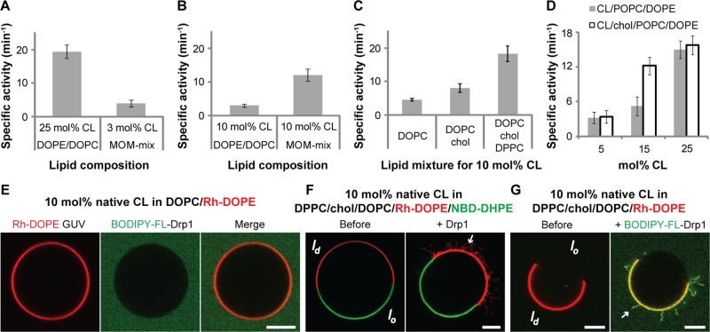
Drp1 preferentially remodels membranes containing a high spatial density of fluid-phase CL. (A–D) Stimulated GTPase activity of Drp1 WT (0.5 μM final) preassembled on liposomes of defined lipid composition (150 μM total lipid) containing varying mole fractions (mol%) of native CL plotted as specific activity (min−1) ± SD (n = 3). (E) Confocal fluorescence images of surface-immobilized Rh-DOPE–labeled GUVs containing 10 mol% native CL (red; left) in the presence of BODIPY-FL-labeled Drp1 WT (0.5 μM protein final; green; middle). Right, merged images. (F) Confocal fluorescence images of GUVs phase-separated into raft-phase, liquid-ordered (lo, NBD-DHPE–labeled, green), and fluid-phase, liquid-disordered (ld, Rh-DOPE–labeled, red) membrane regions before (left) and after (right) addition of unlabeled Drp1 WT (0.5 μM final). Only merged images are shown. Arrow points to Drp1-generated membrane tubules originated from Rh-DOPE–labeled, fluid-phase membrane regions. (G) Same as F, but containing unlabeled, dark, raft-phase lo regions before (left) and after (right) addition of BODIPY-FL–labeled Drp1 WT. Arrow points to Drp1-decorated membrane tubules originating from fluid-phase membrane regions. Only merged images are shown. Scale bar, 5 μm.

