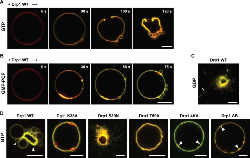
FIGURE 6:
GTP-dependent apparent CL phase transition causes membrane constriction. (A) Time course of Rh-DOPE–labeled GUV membrane morphology changes upon BODIPY-FL–labeled Drp1 WT (0.5 μM final) addition in the constant presence of GTP (1 mM). (B) Same as A, but in the constant presence of GMP-PCP. (C) Endpoint confocal fluorescence image of Drp1 WT-induced GUV membrane remodeling as before but in the constant presence of GDP. (D) same as C, but for various Drp1 mutants in the constant presence of GTP. Slender arrow in C points to Drp1-decorated membrane tubules. In the D panels, slender arrows point to constricted “membrane buds,” whereas triangular, block arrows point to condensed regions of the membrane that presumably mark sites of extensive CL lamellar-to-HII phase transition. Scale bar, 5 μm.

