Figure 3.
Ectopic expression of G2/M-specific genes in proliferating and post-mitotic quiescent cells upon loss of repressor MYBs
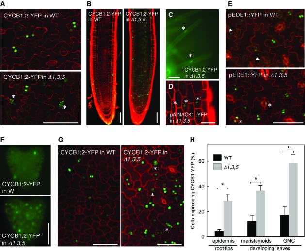
- Expression of CYCB1;2-YFP in the leaf epidermis of 9-day-old plants. Leaves from myb3r1/3/5 (Δ1,3,5) and wild-type (WT) plants were counterstained with propidium iodide for cell walls to visualize cell outlines and were analyzed by confocal microscopy. In myb3r1/3/5 leaves, CYCB1;2-YFP expression was often observed in cells with enlarged nuclei that had presumably undergone endoreduplication, as indicated by asterisks.
- Expression of CYCB1;2-YFP in roots of 3-day-old seedlings. CYCB1;2-YFP expression was expanded toward the basal zone of roots in myb3r1/3/5 seedlings.
- Ectopic expression of CYCB1;2-YFP in terminally differentiated root hair cells in myb3r1/3/5 seedlings (asterisks).
- Expression of proAtNACK1::YFP in epidermal non-dividing cells in myb3r1/3/5 hypocotyl (asterisks).
- Ectopic expression of proEDE1::YFP in maturing guard cells in myb3r1/3/5 leaves (asterisks). Such expression was absent in wild-type leaves (arrowheads).
- Expression of CYCB1;2-YFP in the developing embryo. In a myb3r1/3/5 embryo, a greater population of cells expressed CYCB1;2-YFP compared with a wild-type embryo.
- Expression of CYCB1;2-YFP in cotyledon from 3-day-old seedlings. Asterisks indicate YFP expression in endoreduplicated cells with enlarged nuclei.
- Quantitative comparison of CYCB1;2-YFP-expressing cells in myb3r1/3/5 and wild-type plants. Proportion of CYCB1;2-YFP-expressing cells was determined among epidermal cells in root tips of 5-day-old seedlings (n = 12), and meristemoids and guard mother cells (GMC) in first leaf pairs of 9-day-old seedlings (n = 8). Error bars represent SD. The asterisks in the graphs show differences that are statistically significant (t-test P-value < 0.05).
Data Information: WT, wild type; Δ1,3,5, myb3r1/3/5 triple mutant. Scale bars, 50 μm in (A–E, G), 200 μm in (F).

