
Figure 2.
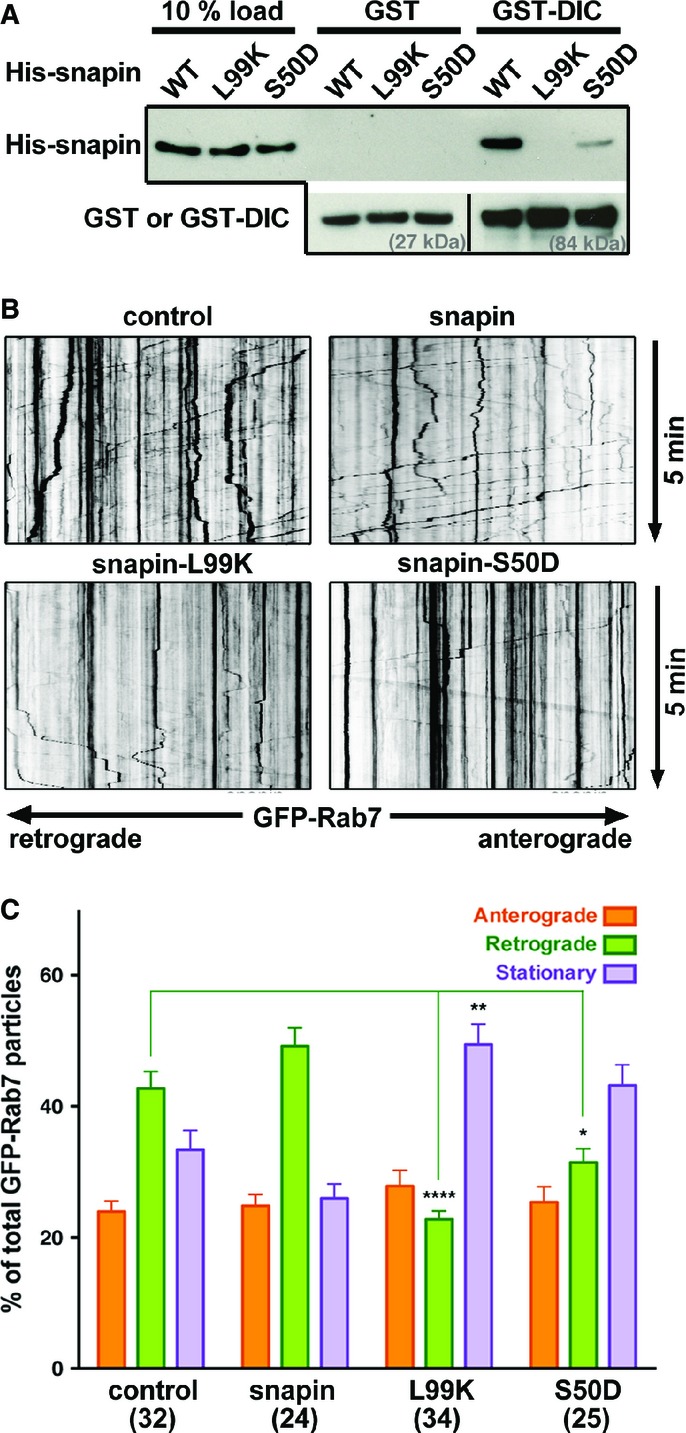
Snapin mutants disturb LE retrograde transport in axons
- A Representative GST-DIC pull-down illustrating distinct binding capacities of WT and mutant snapin with dynein intermediate chain (DIC). Note that the L99K mutation abolished snapin binding to DIC and the S50D mutation robustly decreased snapin–DIC coupling. The same membranes were sequentially blotted with anti-His and anti-GST antibodies. Lower panels demonstrate similar amount of GST or GST-tagged DIC used for the pull-down.
- B, C Representative kymographs (B) and quantitative analysis (C) showing the relative motility of axonal LEs during 5-min time-lapse imaging from cortical neurons at DIV14. Neurons were co-transfected with GFP-Rab7 and a pcDNA vector alone as a control or expressing WT or mutant snapin as indicated. Vertical lines correspond to stationary organelles; oblique traces reflect retrograde (leftward) or anterograde (rightward) transport. Motile or stationary organelles were normalized to the total number of organelles per axon segment selected for recording and averaged from the total number of axons indicated in parentheses. Retrograde transport of Rab7-labeled LEs was robustly inhibited by expressing snapin-L99K and reduced by expressing snapin-S50D. Data are means ± s.e.m., Kruskal–Wallis test with Dunn’s post hoc test. P-values: *0.01–0.05; **0.001 to 0.01; **** < 0.0001. (See also Supplementary Fig S1.)