
Figure 4.
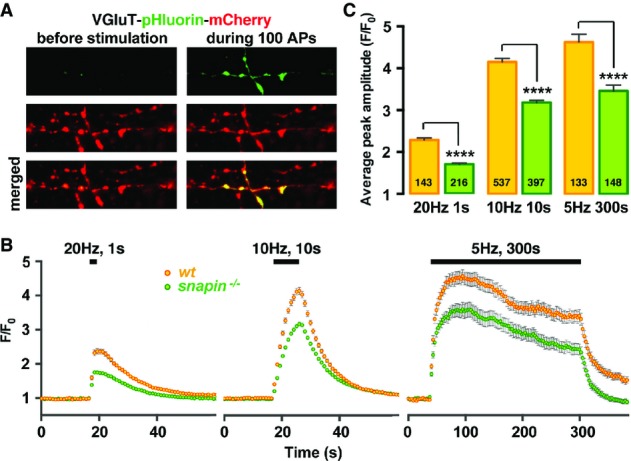
Altered SV exocytosis occurs at snapin-deficient presynaptic terminals
- A Sample pHluorin images from synapses expressing VGluT-pHluorin-mCherry before and during a 10-Hz, 10-s stimulation (100 APs) in cortical neurons at DIV14. Exposure of pHluorin to the alkaline extracellular media during exocytosis gives rise to green fluorescence emission.
- B, C Average pHluorin traces (B) and peak amplitudes (C) elicited by 20 (20 Hz, 1 s), 100 (10 Hz, 10 s), or 1,500 (5 Hz, 300 s) APs. Deleting snapin decreases total releasable pool size without affecting SVs recycling rate during a prolonged (300 s) stimulation. The total number of boutons analyzed is indicated within the columns. Data are means ± s.e.m., Student’s t-test. P-value: **** < 0.0001.