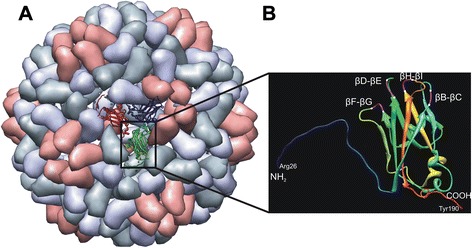
Fig. 1.

Schematical presentation of a CCMV virion and a coat protein subunit. a CCMV particle according to data from Protein Data Bank (PDB ID: 1ZA7 [http://www.rcsb.org/pdb/home/home.do]) [20] and as visualized by Chimera1.6.2 (http://www.cgl.ucsf.edu/chimera/) [60], showing an icosahedral asymmetric unit consisting of three identical subunits in the centre, b ribbon diagram of a coat protein subunit B displaying N-terminal end (residues 1-25 are not shown), four β-barrels (βB-βC, βD-βE, βF-βG and βH-βI) and C-terminal end as potential insertion sites. Insertions within the barrels are shown between the white dashes
