
Figure 5.
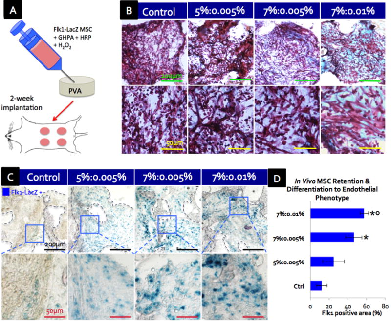
(A) Schematic of in vivo experiment where Flk1-LacZ MSCs-containing GHPA was injected into and crosslinked within a porous PVA scaffold for a murine ventral subcutaneous implantation. (B) Trichrome green staining of cross-sections of scaffolds at 2 weeks post implantation where cytoplasm is stained red, erythrocytes pink and collagen/GHPA gels blue/green. (C) β-galactosidase staining shows that delivered Flk1-LacZ transgenic MSCs were retained and became Flk1-LacZ+(blue) post 2-week implantation in crosslinked GHPA conditions. The boxes indicate Flk1-LacZ+ cell-containing areas. (D) Quantification of retained MSCs that differentiated into an endothelial phenotype (Flk1-LacZ+ cells) post 2-week implantation with error bars = ±1 SEM and N=4. Statistical significance with p < 0.05 is indicated with * in comparison to the control, and ○ in comparison to 5%:0.005%. (B-C) Top row images with scale bars = 200 μm, and bottom row images with scale bars = 50μm.