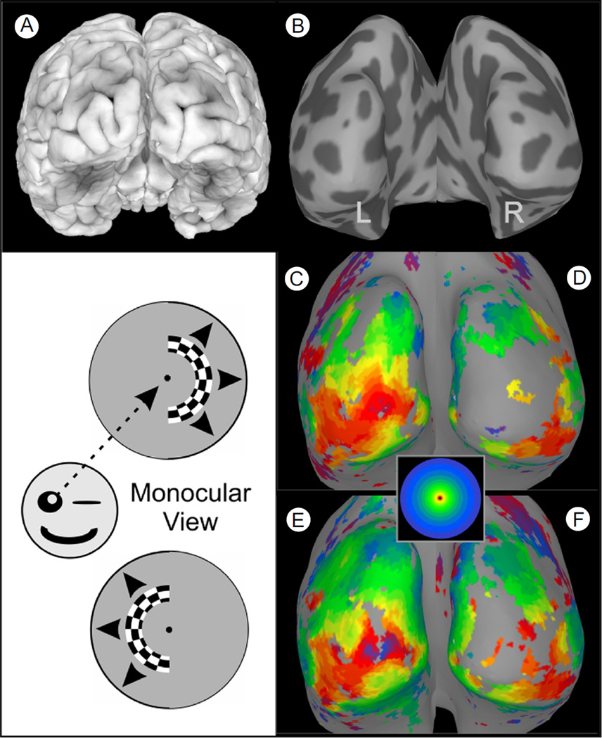
Figure 8.
Aberrant cortical maps in albinism. (A) Posterior view of a 3-dimensional cortical surface model of an albinistic patient's brain. (B) A smoothed cortical surface model obtained by computationally “inflating” the surface model of A. Dark gray patches represent the interior of sulci. (C and D) Left and right hemisphere surface maps showing the pattern of fMRI activation produced when the expanding ring stimulus was confined to the right visual field and viewed monocularly with the right eye (as depicted by the schematic directly to the left). This produced activation that was largely confined to the left hemisphere, contralateral to the stimulus, as would be the case for a nonalbinistic subject. However, when the checkered hemiring was presented in the left visual field (lower schematic), the resulting pattern of activation was, again, strongest and most complete in the left hemisphere (now ipsilateral to the stimulus). Thus, for an albinistic patient, opposite halves of the visual field projected onto the same, left occipital cortex. Note: The aberrant mapping in albinism is not typically apparent if the stimuli are viewed binocularly, because the opposite eye “fills in” the pattern for the opposite hemisphere. (Color version of figure is available online.)

