
Fig. 2. Impaired spermatogenesis in the Xpa (−/−) mice.
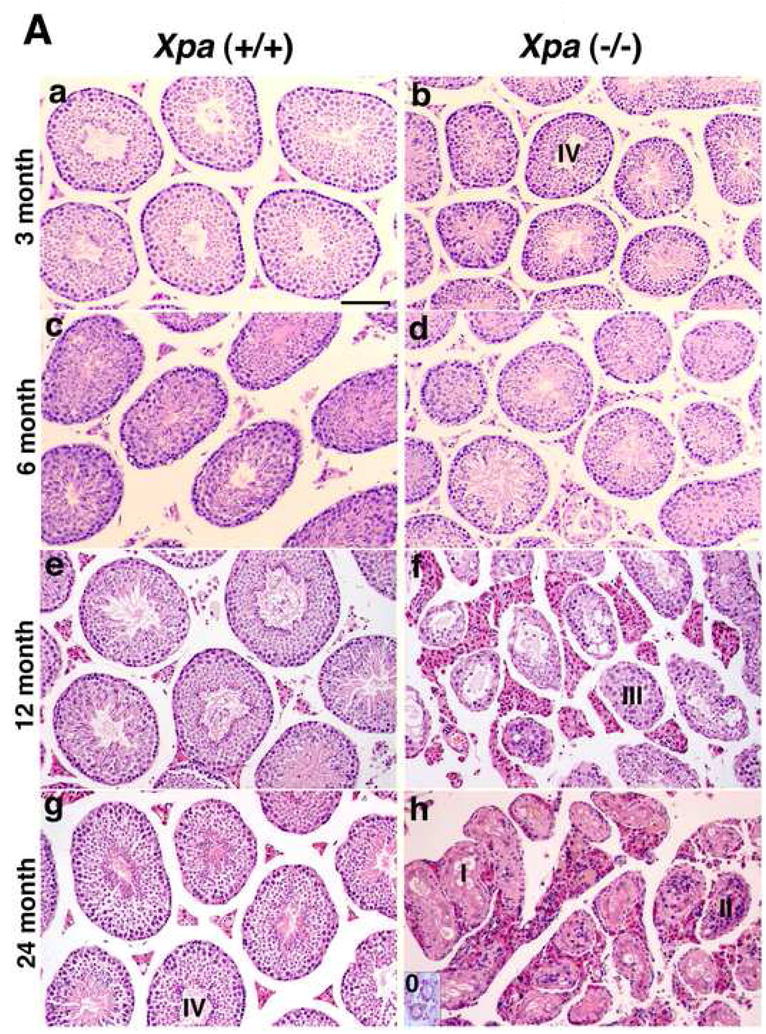
(A). Histology of testis in the Xpa (+/+) and Xpa (−/−) mice.
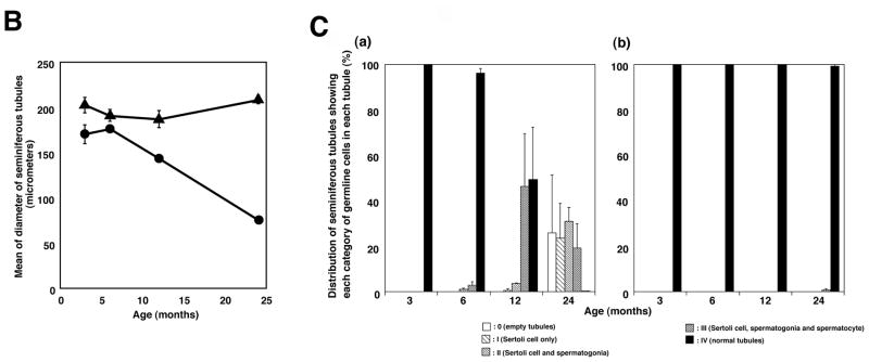
Histological sections were prepared as described in the Materials and methods 2.3. and stained with hematoxylin and eosin. The panels to the left (a, c, e, g) show the testis of Xpa (+/+) mice, while the panels to the right (b, d, f, h) show that of Xpa (−/−) mice. (a), (b): 3-month-old mice; (c), (d): 6-month-old mice; (e), (f): 12-month-old mice; (g), (h): 24-month-old mice. Bar in (a) corresponds to 100μm. Seminiferous tubules were separated into 5 categories (see Materials and methods 2.3). 0-Empty tubules, I-Sertoli cells only, II-Sertoli cells and spermatogonia, III- Sertoli cells, spermatogonia, and spermatocytes, and IV-normal tubules. Note that in (a) and (b), the diameter of seminiferous tubules in the 3-month-old Xpa (−/−) mice is shorter than in the Xpa (+/+) littermates; in (d), the diameters of seminiferous tubules in the Xpa (−/−) mice are very variable; in (f), the testis displayed grossly dysmorphic seminiferous tubules (vacuolated tubules, disarrays of germ cells) with hyperplasia of Leydig cells; in (h), the testis displayed completely disrupted seminiferous tubules, and empty tubules were shown in the inset of Fig. 2Ah. (B). The diameter of seminiferous tubules in the Xpa (+/+) and Xpa (−/−) mice. The mean and SEM of diameters are indicated. Closed triangle: Xpa (+/+) mice; closed circle: Xpa (−/−) mice. Note that the diameter of seminiferous tubules in the Xpa (−/−) mice decreased in an age-dependent manner. (C). Classification of the seminiferous tubules into 5 categories depending on the germline cells and the chronological distribution of each category. (a) testis of Xpa (−/−) mice; (b) testis of Xpa (+/+) mice, □ category 0 (empty tubules) [Fig. 2A, h, inset];  category I (Sertoli cells only) [Fig. 2A, h];
  category II (Sertoli cells and spermatogonia) [Fig. 2A, h];
  category III (Sertoli cells, spermatogonia, and spermatocytes) [Fig. 2A, f]; ■ category IV (normal tubules) [Fig. 2Ab, g]. The mean value and SEM of the frequency of each category are indicated. Degenerative seminiferous tubules (category 0, I, II, and III) gradually increased in the Xpa (−/−) mice in an age-dependent manner. For the classification analysis, mice without tumors were examined.