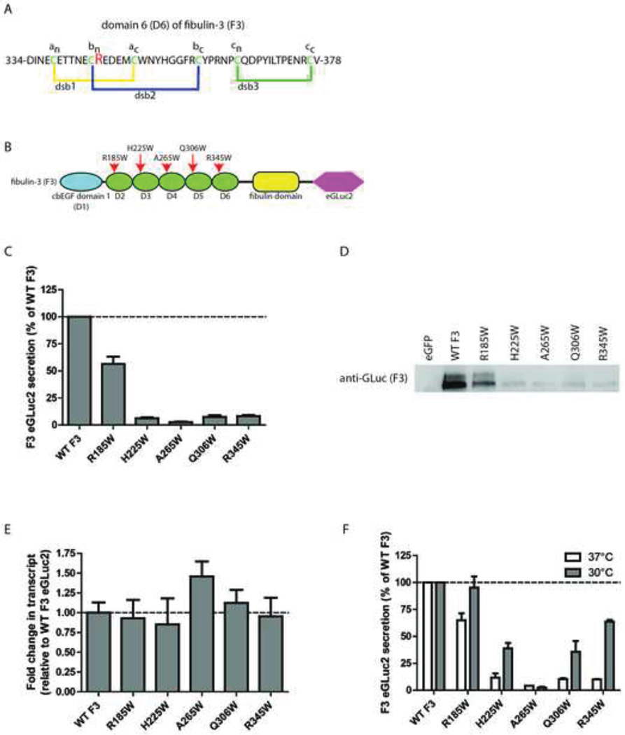
Fig. 1.
Introduction of tryptophan residues into cbEGF domains of F3 have distinct effects on F3 secretion. (A) Schematic representation of domain 6 (D6) of F3, adapted from (Hulleman et al., 2011). dsb = disulfide bond (B) F3 domain organization and location of newly generated tryptophan residues, adapted from (Hulleman et al., 2011). (C) Conditioned media aliquots from transfected TREx-293 cells were taken 48 h after transfection and assayed for GLuc activity. (D) Western blot verification of GLuc activity levels shown in (C). (E) qPCR verification of similar F3 expression levels. mRNA was isolated from TREx-293 cells 48 h after transfection and the transcript levels of F3 relative to WT F3 were evaluated by qPCR. (F) Transfected TREx-293 cells were grown at 37°C followed by a media change and 24 h at 30°C. Conditioned media aliquots were assayed using the GLuc assay. n ≥ 3 independent experiments for panels C–F.

