
Fig. 3.
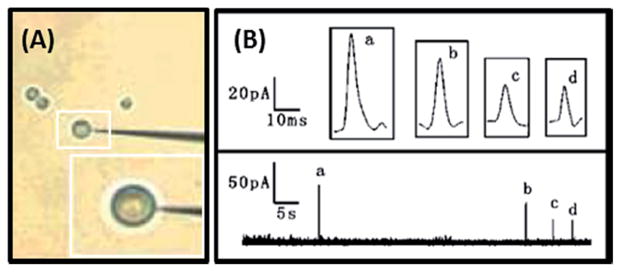
(A) Photograph of nanoelectrode-cell arrangement. (B) Spikes corresponding to sequential dopamine release from multiple vesicles with the electrode placed over the same position on the cell. Graphs a–d show magnified pictures of the individual current spikes. Modified from ref. 77.