Figure 1.

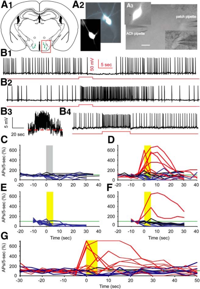
Cholinergic stimulation by ACh boosts spontaneous action potential firing in Hcrt+ neurons. A1, Sketch of a brain slice showing Hcrt+ neurons (green cells in the red box) residing in the hypothalamus. A2, Morphology of two Hcrt+ neurons shown in the fluorescent channel. A3, Differential Interference Contrast video-microscopy showing the experimental paradigm of pressure application (puff) of drug onto the soma and proximal processes, while keeping the patch onto the neuron. Scale bar, 10 µm. B1, Mechanical interference (puff of bath solution) frequently produces a temporary depression of spontaneous firing. B2, In the presence of atropine (4 µM), application of ACh (1 mM) boosts action potential firing for tens of seconds. B3, The same trace as in B2, except on a different scale and filtered with Gaussian low pass 5 Hz. B4, Injection of +10 pA of current into the Hcrt+ neuron notably increases the firing frequency. Red lines represent the timing and duration of ACh application or current injection. C, Mechanical interference does not affect firing (n = 4/11 cells), or results in a temporary depression of firing (n = 7/11 cells). D, Differential responses to the puff of ACh. In 7 out of 20 cells, firing was enhanced by ACh, 4 out of 20 cells were not affected, while 9 out of 20 cells were inhibited. E, With DHβE in the bath, ACh did not increase firing of any Hcrt+ neurons tested (n = 11/11 cells). F, With MLA in the bath, a puff of ACh boosted firing in 2 of 16 cells, had no effect in 9 of 16 cells, and decreased firing in 5 of 16 cells. The gray bar indicates the time duration of ACSF application; yellow bars indicate the time duration of ACh application. C−F, All experiments were conducted in the presence of atropine. G, The responses of Hcrt+ neurons to ACh in absence of atropine. In 8 out of 14 cells, firing was enhanced by ACh, in 1 out of 14 cells firing was unaffected, while in 5 out of 14 cells firing was inhibited.
